Choroidal arteries: Difference between revisions
Jump to navigation
Jump to search
| (6 intermediate revisions by the same user not shown) | |||
| Line 1: | Line 1: | ||
= Anterior Choroidal arteries = | |||
{{NoteBox|primary}}<small>ant. choroidal a.: takeoff 2–4mm distal to PComA ⇒ (variable) portion of optic tract, medial GP, genu of IC (in 50%), inf. half of pos. limb of IC, uncus, retrolenticular fibers (optic radiation), LGBN; <strong>AChA syndrome</strong>, triad: CL hemiplegia, hemihypesthesia & homonymous hemianopsia (<mark>mnemonic: 3 H’s</mark>); however, incomplete forms are MC; Occlusion is usually d/t small vessel dz → pos. limb of IC</small>{{NoteBoxEnd}} | |||
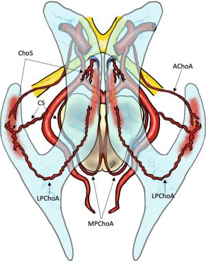
[[File:Embryology and Variations of the Posterior Choroidal Artery.jpg|alt=Embryology and Variations of the Posterior Choroidal Artery|thumb|Embryology and Variations of the Posterior Choroidal Artery]] | [[File:Embryology and Variations of the Posterior Choroidal Artery.jpg|alt=Embryology and Variations of the Posterior Choroidal Artery|thumb|Embryology and Variations of the Posterior Choroidal Artery]] | ||
* Arises from the posteromedial surface of the [[ICA]], 2-4 mm distal to the Pcomm origin. | |||
* | * It anastomoses with the lateral posterior ChAs and has two segments | ||
* Course: goes through the choroidal fissure → temporal horn of LV | * Course: goes through the choroidal fissure → temporal horn of LV | ||
* Historically, this artery was sacrificed to treat Parkinson’s disease: decreased tremor likely due to decreased blood supply to VL thalamus. | * Historically, this artery was sacrificed to treat Parkinson’s disease: decreased tremor likely due to decreased blood supply to VL thalamus. | ||
== Segments of AChA == | |||
* <strong>Cisternal segment</strong> - courses posteromedially within the suprasellar cistern beneath (inferior and lateral) the optic tract, then turns posteromedially around the uncus | |||
* <strong>Intraventricular segment</strong> - continues from the cisternal segment. Prior to reaching the lateral geniculate body, it turns posterolaterally through the crural and ambient cisterns to enter the choroidal fissure (plexal point) of the temporal horn. | |||
== Vascular territory of AChA == | |||
Perforators (3-10) supply: | |||
* Visual system - inferior optic chiasm, posterior portion of the optic tract, optic radiation, lateral geniculate body | |||
* Temporal lobe - uncus, hippocampus/parahippocampal gyrus, amygdala | |||
* Choroid plexus in the temporal horn (lateral horn) and atrium of the LVs | |||
* Basal ganglia - globus pallidus medius, tail of caudate, internal capsule (genu, posterior limb and retrolenticular) | |||
* Diencephalon - subthalamus, lateral ventroanterior (VA) and ventrolateral (VL) thalamic nuclei | |||
* Midbrain — middle third of the cerebral peduncle, upper red nucleus, substantia nigra | |||
= Posterior Choroidal arteries (medial and lateral) = | = Posterior Choroidal arteries (medial and lateral) = | ||
Latest revision as of 23:32, 4 March 2024
Anterior Choroidal arteries
ant. choroidal a.: takeoff 2–4mm distal to PComA ⇒ (variable) portion of optic tract, medial GP, genu of IC (in 50%), inf. half of pos. limb of IC, uncus, retrolenticular fibers (optic radiation), LGBN; AChA syndrome, triad: CL hemiplegia, hemihypesthesia & homonymous hemianopsia (mnemonic: 3 H’s); however, incomplete forms are MC; Occlusion is usually d/t small vessel dz → pos. limb of IC

- Arises from the posteromedial surface of the ICA, 2-4 mm distal to the Pcomm origin.
- It anastomoses with the lateral posterior ChAs and has two segments
- Course: goes through the choroidal fissure → temporal horn of LV
- Historically, this artery was sacrificed to treat Parkinson’s disease: decreased tremor likely due to decreased blood supply to VL thalamus.
Segments of AChA
- Cisternal segment - courses posteromedially within the suprasellar cistern beneath (inferior and lateral) the optic tract, then turns posteromedially around the uncus
- Intraventricular segment - continues from the cisternal segment. Prior to reaching the lateral geniculate body, it turns posterolaterally through the crural and ambient cisterns to enter the choroidal fissure (plexal point) of the temporal horn.
Vascular territory of AChA
Perforators (3-10) supply:
- Visual system - inferior optic chiasm, posterior portion of the optic tract, optic radiation, lateral geniculate body
- Temporal lobe - uncus, hippocampus/parahippocampal gyrus, amygdala
- Choroid plexus in the temporal horn (lateral horn) and atrium of the LVs
- Basal ganglia - globus pallidus medius, tail of caudate, internal capsule (genu, posterior limb and retrolenticular)
- Diencephalon - subthalamus, lateral ventroanterior (VA) and ventrolateral (VL) thalamic nuclei
- Midbrain — middle third of the cerebral peduncle, upper red nucleus, substantia nigra
Posterior Choroidal arteries (medial and lateral)
Medial Posterior Choroidal Artery

- Medial branches supply pineal gland, tectum, thalamus, choroid of the third ventricle
- Usually arise from the proximal P2 of the PCA
- הולך אחורה בציסטרנה אמביאנס
- בהמשך מסתובב קדימה לתוך גג החדר השלישי
Lateral Posterior Choroidal Artery
- Lateral branches enter the choroidal fissure and anastomose with the anterior choroidal arteries forming variable anastomotic network