Posterior Cerebral Artery: Difference between revisions
Jump to navigation
Jump to search
No edit summary |
|||
| (15 intermediate revisions by the same user not shown) | |||
| Line 1: | Line 1: | ||
{{NoteBox|secondary}}<strong>Related pages</strong> | |||
* [[Fetal PCA]] | |||
{{NoteBoxEnd}} | |||
<br clear="all"> | |||
= Vascular territory = | |||
* Parietooccipital sulcus (medial) and inferior temporal sulcus (lateral) | |||
= Anatomical segments = | = Anatomical segments = | ||
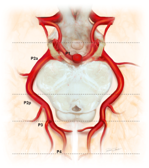
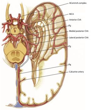
* P1: PCA from the origin to posterior communicating artery (AKA mesencephalic, precommunicating, circular, peduncular, basilar...). The long and short circumflex and thalamoperforating arteries arise from P1 | [[File:PCA Segments.png|thumb]] | ||
* P2: PCA from origin of PComA to the origin of inferior temporal arteries (AKA ambient, post-communicating, perimesencephalic), P2 traverses the ambient cistern, hippocampal, anterior temporal, peduncular perforating, and medial posterior choroidal arteries arise from P2 | * P1: PCA from the origin to [[Posterior Communicating Artery|posterior communicating artery]] (AKA mesencephalic, precommunicating, circular, peduncular, basilar...). The long and short circumflex and thalamoperforating arteries arise from P1 | ||
* P2: PCA from origin of [[PComA]] to the origin of inferior temporal arteries (AKA ambient, post-communicating, perimesencephalic), P2 traverses the ambient cistern, hippocampal, anterior temporal, peduncular perforating, and medial posterior [[choroidal arteries]] arise from P2 | |||
* P3: PCA from the origin of the inferior temporal branches to the origin of the terminal branches (AKA quadrigeminal segment). P3 traverses the quadrigeminal cistern | * P3: PCA from the origin of the inferior temporal branches to the origin of the terminal branches (AKA quadrigeminal segment). P3 traverses the quadrigeminal cistern | ||
* P4: segment after the origin of the parieto-occipital and calcarine arteries, includes the cortical branches of the PCA | * P4: segment after the origin of the parieto-occipital and calcarine arteries, includes the cortical branches of the PCA | ||
[[Category:Neuroanatomy]] | <br clear="all"> | ||
[[Category:Vascular anatomy]] | {| class="wikitable" | ||
|- | |||
! style="width: 30%; vertical-align: top;" | Segment | |||
! style="width: 70%; vertical-align: top;" | Description | |||
|- | |||
| style="vertical-align: top;" | P1 | |||
| style="vertical-align: top;" | Arises from the basilar bifurcation and extends through the interpeduncular cistern to the junction with the [[Posterior Communicating Artery|posterior communicating artery]] | |||
|- | |||
| style="vertical-align: top;" | P2A (anterior) | |||
| style="vertical-align: top;" | Runs in the crural cistern, extending from the PComA to the lateral mesencephalic sulcus where it becomes the P2P (posterior) segment | |||
|- | |||
| style="vertical-align: top;" | P2P (posterior) | |||
| style="vertical-align: top;" | Runs in the ambient cistern, lateral to the midbrain | |||
|- | |||
| style="vertical-align: top;" | P3 | |||
| style="vertical-align: top;" | Runs in the quadrigeminal cistern, separated from the P2P segment by the tectal plate | |||
|- | |||
| style="vertical-align: top;" | P4 | |||
| style="vertical-align: top;" | Begins at the calcarine sulcus | |||
|} | |||
[[File:Posterior cerebral artery.jpg|thumb]] | |||
= Branches of PCA = | |||
== P1 segment == | |||
<table class="wikitable" width="100%"> | |||
<tr> | |||
<th width="30%">P1 Segment</th> | |||
<th>Feature</th> | |||
</tr> | |||
<tr> | |||
<td>Posterior thalamoperforator arteries</td> | |||
<td> | |||
* From the basilar artery and P1, travels through the posterior perforated substance behind the mamillary bodies | |||
* Supply: thalamus, hypothalamus, subthalamus, and midbrain ([[cranial nerves]] III and IV)</td> | |||
</tr> | |||
<tr> | |||
<td>[[Choroidal arteries|Medial posterior choroidal arteries]]</td> | |||
<td> | |||
* Travels anteromedially along the roof of the third ventricle | |||
* Supply: midbrain tectum, posterior thalamus, pineal gland, and tela choroidae of the third ventricle</td> | |||
</tr> | |||
<tr> | |||
<td>Meningeal branches</td> | |||
<td>Supply: tentorium and the falx</td> | |||
</tr> | |||
</table> | |||
== P2 segment == | |||
<table class="wikitable" width="100%"> | |||
<tr> | |||
<th width="30%">P2 Segment</th> | |||
<th>Feature</th> | |||
</tr> | |||
<tr> | |||
<td>[[Choroidal arteries|Lateral posterior choroidal artery]]</td> | |||
<td> | |||
* Main branch of P2, courses over the pulvinar and through the choroidal fissure | |||
* Supply: posterior portion of the thalamus and choroid plexus (temporal horn and atrium) | |||
</td> | |||
</tr> | |||
<tr> | |||
<td>Thalamogeniculate arteries</td> | |||
<td>Medial geniculate body, lateral geniculate body, pulvinar, superior colliculus, and crus cerebri</td> | |||
</tr> | |||
<tr> | |||
<td>Cortical branches</td> | |||
<td> | |||
* Inferior temporal artery group | |||
* Supply: inferior portion of the temporal lobe | |||
</td> | |||
</tr> | |||
</table> | |||
== P3 segment == | |||
<table class="wikitable" width="100%"> | |||
<tr> | |||
<th width="30%">P3 Segment</th> | |||
<th>Feature</th> | |||
</tr> | |||
<tr> | |||
<td>Posterior temporal artery</td> | |||
<td> | |||
* Posterior temporal lobe, occipitotemporal and lingual gyri | |||
* Anterior temporal artery branch travels to the inferior temporal lobe to supply the inferior cortex. | |||
* Anastomoses with middle cerebral artery | |||
</tr> | |||
<tr> | |||
<td>Internal occipital artery</td></tr><tr> | |||
<td>Parietooccipital artery</td> | |||
<td> | |||
* Located in the parietooccipital sulcus | |||
* Supply: posterior ⅓ of the medial hemispheres, cuneus, precuneus, superior occipital gyrus, and precentral and superior parietal lobules | |||
* Anastomoses with anterior cerebral artery | |||
</td> | |||
</tr> | |||
<tr> | |||
<td>Calcarine artery</td> | |||
<td> | |||
* Located in the calcarine sulcus | |||
* Supply: occipital pole and the visual cortex | |||
* Anastomoses with middle cerebral artery | |||
</td> | |||
</tr> | |||
<tr> | |||
<td>Posterior pericallosal artery</td> | |||
<td> | |||
* Supply: splenium of the corpus callosum | |||
* Anastomoses with anterior cerebral artery</td> | |||
</tr> | |||
</table> | |||
[[index.php?title=Category:Neuroanatomy]] | |||
[[index.php?title=Category:Vascular anatomy]] | |||
Latest revision as of 06:33, 3 August 2024
Related pages
Vascular territory
- Parietooccipital sulcus (medial) and inferior temporal sulcus (lateral)
Anatomical segments

- P1: PCA from the origin to posterior communicating artery (AKA mesencephalic, precommunicating, circular, peduncular, basilar...). The long and short circumflex and thalamoperforating arteries arise from P1
- P2: PCA from origin of PComA to the origin of inferior temporal arteries (AKA ambient, post-communicating, perimesencephalic), P2 traverses the ambient cistern, hippocampal, anterior temporal, peduncular perforating, and medial posterior choroidal arteries arise from P2
- P3: PCA from the origin of the inferior temporal branches to the origin of the terminal branches (AKA quadrigeminal segment). P3 traverses the quadrigeminal cistern
- P4: segment after the origin of the parieto-occipital and calcarine arteries, includes the cortical branches of the PCA
| Segment | Description |
|---|---|
| P1 | Arises from the basilar bifurcation and extends through the interpeduncular cistern to the junction with the posterior communicating artery |
| P2A (anterior) | Runs in the crural cistern, extending from the PComA to the lateral mesencephalic sulcus where it becomes the P2P (posterior) segment |
| P2P (posterior) | Runs in the ambient cistern, lateral to the midbrain |
| P3 | Runs in the quadrigeminal cistern, separated from the P2P segment by the tectal plate |
| P4 | Begins at the calcarine sulcus |

Branches of PCA
P1 segment
| P1 Segment | Feature |
|---|---|
| Posterior thalamoperforator arteries |
|
| Medial posterior choroidal arteries |
|
| Meningeal branches | Supply: tentorium and the falx |
P2 segment
| P2 Segment | Feature |
|---|---|
| Lateral posterior choroidal artery |
|
| Thalamogeniculate arteries | Medial geniculate body, lateral geniculate body, pulvinar, superior colliculus, and crus cerebri |
| Cortical branches |
|
P3 segment
| P3 Segment | Feature |
|---|---|
| Posterior temporal artery |
|
| Internal occipital artery | |
| Parietooccipital artery |
|
| Calcarine artery |
|
| Posterior pericallosal artery |
|
index.php?title=Category:Neuroanatomy index.php?title=Category:Vascular anatomy