Facial Nerve: Difference between revisions
Jump to navigation
Jump to search
No edit summary |
m (The LinkTitles extension automatically added links to existing pages (https://github.com/bovender/LinkTitles).) |
||
| (8 intermediate revisions by 2 users not shown) | |||
| Line 1: | Line 1: | ||
== Clinical grading of facial nerve function (House and Brackmann) == | == Anatomy and Course == | ||
* The facial nerve (Cranial Nerve VII) is a mixed nerve that controls the muscles of facial expression and conveys taste sensations from the anterior two-thirds of the tongue. | |||
* It originates in the pons and exits the [[brainstem]] at the cerebellopontine angle. | |||
* The nerve then enters the [[internal auditory canal]], runs through the facial canal in the temporal bone, and exits the skull via the stylomastoid foramen. | |||
* Within the temporal bone, the facial nerve gives off the greater petrosal nerve, nerve to stapedius, and chorda tympani. | |||
== Nuclei of the Facial Nerve == | |||
=== Motor Nucleus: === | |||
* Located in the pons. | |||
* Controls muscles of facial expression. | |||
=== Superior Salivatory Nucleus: === | |||
* Provides parasympathetic innervation to the lacrimal, nasal, and palatine glands. | |||
=== Nucleus of the Solitary Tract: === | |||
* Receives taste sensations from the anterior two-thirds of the tongue. | |||
=== Spinal Trigeminal Nucleus: === | |||
* Processes pain and temperature sensations from the ear. | |||
== Segments of the Facial Nerve == | |||
{| class="wikitable" | |||
|'''Segment''' | |||
|'''Anatomy''' | |||
|'''Symptoms''' | |||
|'''Clinical Significance''' | |||
|- | |||
|Intracranial (Cisternal) Segment | |||
|Extends from the brainstem to the internal auditory meatus. | |||
| | |||
* Complete facial paralysis on the affected side. | |||
* Loss of taste sensation from the anterior two-thirds of the tongue. | |||
* Decreased lacrimation and salivation. | |||
|These symptoms might be accompanied by other cranial nerve deficits if the cause is a cerebellopontine angle tumor or similar pathology. | |||
|- | |||
|Meatal (Labyrinthine) Segment | |||
|Runs within the internal auditory canal. | |||
| | |||
* Facial paralysis, including the forehead (frontalis muscle). | |||
* Hearing loss or tinnitus if the vestibulocochlear nerve is also affected. | |||
|Commonly seen in acoustic neuroma or during vestibular [[schwannoma]] surgery. | |||
|- | |||
|Tympanic (Horizontal) Segment | |||
|Lies within the temporal bone. | |||
| | |||
* Facial paralysis. | |||
* Hyperacusis (increased sensitivity to certain frequencies and volume ranges of sound) due to stapedius muscle paralysis. | |||
|Middle ear pathologies like cholesteatoma or chronic infection can affect this segment. | |||
|- | |||
|Mastoid (Vertical) Segment | |||
|Descends in the mastoid bone. | |||
| | |||
* Facial paralysis. | |||
* Possible alteration in taste sensation. | |||
|Mastoid surgeries or ear infections can damage this segment. | |||
|- | |||
|Extratemporal Segment | |||
|Emerges from the stylomastoid foramen and branches in the face. | |||
| | |||
* Paralysis of the muscles of facial expression on the affected side, often sparing the forehead in cases of partial damage due to dual innervation. | |||
* If the main trunk is affected, all facial expressions are impaired. | |||
|Commonly injured in facial trauma or parotid gland surgery. | |||
|} | |||
== Terminal Branches and Innervation == | |||
* After exiting the stylomastoid foramen, it branches into five main branches: Temporal, Zygomatic, Buccal, Marginal Mandibular, and Cervical. | |||
* These branches innervate the muscles of facial expression, including the muscles of the forehead, eye, cheek, and neck. | |||
== Functional Aspects == | |||
* The nerve is responsible for facial expressions, eyelid closing (orbicularis oculi muscle), and lip movement. | |||
* It also innervates the stapedius muscle in the middle ear (impacting hearing) and conveys taste sensation from the anterior tongue. | |||
== Clinical Significance == | |||
* Pathologies affecting the facial nerve can lead to facial paralysis or paresis, most commonly Bell’s palsy. | |||
* The nerve is at risk during surgical procedures in the parotid region, temporal bone surgeries, and cerebellopontine angle surgeries. | |||
* Preservation of the facial nerve during surgical interventions is paramount to prevent facial asymmetry and functional impairments. | |||
=== Clinical grading of facial nerve function (House and Brackmann) === | |||
{| class="wikitable" | {| class="wikitable" | ||
!Grade | !Grade | ||
| Line 12: | Line 98: | ||
|'''mild dysfunction''' | |'''mild dysfunction''' | ||
| | | | ||
# gross: slight weakness noticeable on close inspection; may have very slight synkinesis | #gross: slight weakness noticeable on close inspection; may have very slight synkinesis | ||
# at rest: normal symmetry and tone | #at rest: normal symmetry and tone | ||
# motion: | #motion: | ||
## forehead: slight to moderate movement | ##forehead: slight to moderate movement | ||
## eye: complete closure with effort | ##eye: complete closure with effort | ||
## mouth: slight asymmetry | ##mouth: slight asymmetry | ||
|- | |- | ||
|3 | |3 | ||
|'''moderate dysfunction''' | |'''moderate dysfunction''' | ||
| | | | ||
# gross: obvious but not disfiguring asymmetry; noticeable but not severe synkinesis | #gross: obvious but not disfiguring asymmetry; noticeable but not severe synkinesis | ||
# motion: | #motion: | ||
## forehead: slight to moderate movement | ##forehead: slight to moderate movement | ||
## eye: complete closure with effort | ##eye: complete closure with effort | ||
## mouth: slightly weak with maximal effort | ##mouth: slightly weak with maximal effort | ||
|- | |- | ||
|4 | |4 | ||
|'''moderate to severe dysfunction''' | |'''moderate to severe dysfunction''' | ||
| | | | ||
# gross: obvious weakness and/or disfiguring asymmetry | #gross: obvious weakness and/or disfiguring asymmetry | ||
# motion: | #motion: | ||
## forehead: none | ##forehead: none | ||
## eye: incomplete closure | ##eye: incomplete closure | ||
## mouth: asymmetry with maximum effort | ##mouth: asymmetry with maximum effort | ||
|- | |- | ||
|5 | |5 | ||
|'''severe dysfunction''' | |'''severe dysfunction''' | ||
| | | | ||
# gross: only barely perceptible motion | #gross: only barely perceptible motion | ||
# at rest: asymmetry | #at rest: asymmetry | ||
# motion: | #motion: | ||
## forehead: none | ##forehead: none | ||
## eye: incomplete closure | ##eye: incomplete closure | ||
|- | |- | ||
|6 | |6 | ||
| Line 50: | Line 136: | ||
| | | | ||
|} | |} | ||
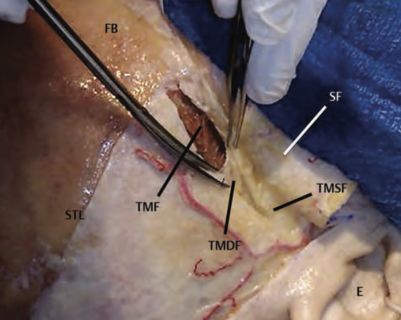
== Surgical Considerations == | |||
* Techniques like interfascial dissection are employed to protect the nerve, especially in procedures involving the scalp and temporal region. | |||
[[File:Preserving frontal branch of the facial nerve through a subfascial dissection.jpg|left|thumb|401x401px|Preserving frontal branch of the facial nerve through a subfascial dissection]] | |||
[[Category:Neuroanatomy]] | |||
[[Category:Cranial nerves]] | |||
Latest revision as of 22:00, 3 March 2024
Anatomy and Course
- The facial nerve (Cranial Nerve VII) is a mixed nerve that controls the muscles of facial expression and conveys taste sensations from the anterior two-thirds of the tongue.
- It originates in the pons and exits the brainstem at the cerebellopontine angle.
- The nerve then enters the internal auditory canal, runs through the facial canal in the temporal bone, and exits the skull via the stylomastoid foramen.
- Within the temporal bone, the facial nerve gives off the greater petrosal nerve, nerve to stapedius, and chorda tympani.
Nuclei of the Facial Nerve
Motor Nucleus:
- Located in the pons.
- Controls muscles of facial expression.
Superior Salivatory Nucleus:
- Provides parasympathetic innervation to the lacrimal, nasal, and palatine glands.
Nucleus of the Solitary Tract:
- Receives taste sensations from the anterior two-thirds of the tongue.
Spinal Trigeminal Nucleus:
- Processes pain and temperature sensations from the ear.
Segments of the Facial Nerve
| Segment | Anatomy | Symptoms | Clinical Significance |
| Intracranial (Cisternal) Segment | Extends from the brainstem to the internal auditory meatus. |
|
These symptoms might be accompanied by other cranial nerve deficits if the cause is a cerebellopontine angle tumor or similar pathology. |
| Meatal (Labyrinthine) Segment | Runs within the internal auditory canal. |
|
Commonly seen in acoustic neuroma or during vestibular schwannoma surgery. |
| Tympanic (Horizontal) Segment | Lies within the temporal bone. |
|
Middle ear pathologies like cholesteatoma or chronic infection can affect this segment. |
| Mastoid (Vertical) Segment | Descends in the mastoid bone. |
|
Mastoid surgeries or ear infections can damage this segment. |
| Extratemporal Segment | Emerges from the stylomastoid foramen and branches in the face. |
|
Commonly injured in facial trauma or parotid gland surgery. |
Terminal Branches and Innervation
- After exiting the stylomastoid foramen, it branches into five main branches: Temporal, Zygomatic, Buccal, Marginal Mandibular, and Cervical.
- These branches innervate the muscles of facial expression, including the muscles of the forehead, eye, cheek, and neck.
Functional Aspects
- The nerve is responsible for facial expressions, eyelid closing (orbicularis oculi muscle), and lip movement.
- It also innervates the stapedius muscle in the middle ear (impacting hearing) and conveys taste sensation from the anterior tongue.
Clinical Significance
- Pathologies affecting the facial nerve can lead to facial paralysis or paresis, most commonly Bell’s palsy.
- The nerve is at risk during surgical procedures in the parotid region, temporal bone surgeries, and cerebellopontine angle surgeries.
- Preservation of the facial nerve during surgical interventions is paramount to prevent facial asymmetry and functional impairments.
Clinical grading of facial nerve function (House and Brackmann)
| Grade | Function Description | Clinical Sx |
|---|---|---|
| 1 | normal facial function in all areas | |
| 2 | mild dysfunction |
|
| 3 | moderate dysfunction |
|
| 4 | moderate to severe dysfunction |
|
| 5 | severe dysfunction |
|
| 6 | total paralysis no movement |
Surgical Considerations
- Techniques like interfascial dissection are employed to protect the nerve, especially in procedures involving the scalp and temporal region.
