Meningioma: Difference between revisions
Jump to navigation
Jump to search
| (13 intermediate revisions by the same user not shown) | |||
| Line 1: | Line 1: | ||
{{NoteBox|secondary}}<strong>Related pages</strong> | {{NoteBox|secondary}}<strong>Related pages</strong> | ||
* [[cavernous sinus meningioma|Cavernous sinus Meningiomas]]{{NoteBoxEnd}} | * [[Anterior skull base Meningiomas]] | ||
* [[cavernous sinus meningioma|Cavernous sinus Meningiomas]] | |||
* [[Parasagittal and falx meningiomas]]{{NoteBoxEnd}} | |||
[[File:BT SupTentPostFossa OlfGrve 02.jpg|thumb|454x454px]] | [[File:BT SupTentPostFossa OlfGrve 02.jpg|thumb|454x454px]] | ||
= General information = | = General information = | ||
| Line 7: | Line 9: | ||
<br clear="all"> | <br clear="all"> | ||
= Diagnosis = | = Diagnosis = | ||
| Line 74: | Line 19: | ||
* Pneumosinus dilatans | * Pneumosinus dilatans | ||
* MRI: isointense on T1WI, hypointense on T2WI | * MRI: isointense on T1WI, hypointense on T2WI | ||
== Histopathology == | |||
[[File:Meningioma - Histopathology .jpg|thumb|500x500px]] | |||
* The tumour cells have features of both syncytial and fibroblastic type forming <mark>whorls</mark> which contain central laminated areas of calcification called <mark>psammoma bodies</mark>. | |||
<br clear="all"> | |||
=== Meningioma Grades === | |||
{| class="wikitable" style="width:100%;" | |||
! GRADE I | |||
! GRADE II | |||
! GRADE III | |||
|- | |||
| | |||
* Meningothelial meningioma | |||
* Fibrous meningioma | |||
* Transitional meningioma | |||
* Psammomatous meningioma | |||
* Angiomatous meningioma | |||
* Microcystic meningioma | |||
* Secretory meningioma | |||
* Lymphoplasmacyte-rich meningioma | |||
* Metaplastic meningioma | |||
| | |||
* Atypical meningioma | |||
* Clear cell meningioma | |||
* Chordoid meningioma | |||
| | |||
* Rhabdoid meningioma | |||
* Papillary meningioma | |||
* Anaplastic (malignant) meningioma | |||
|} | |||
== Immunohistochemical staining == | |||
* EMA ⊕ in ~80% | |||
[[Category:Neuro-Oncology]] | [[Category:Neuro-Oncology]] | ||
[[Category:Meningioma]] | [[Category:Meningioma]] | ||
[[Category:Extrinsic Brain Tumors]] | [[Category:Extrinsic Brain Tumors]] | ||
Latest revision as of 22:57, 4 March 2024
Related pages

General information
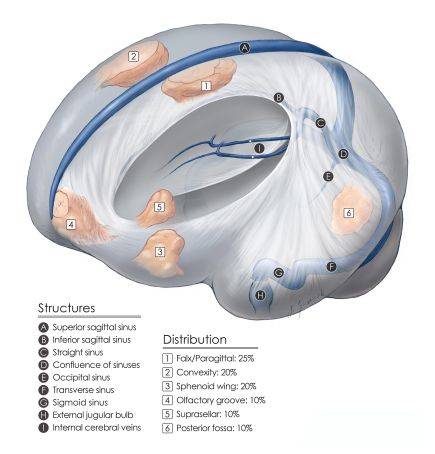
- Meningiomas are a group of tumors that are believed to originate in meningothelial cells of the arachnoid membrane.
- They may be intracranial (MC), intraorbital, or intra-spinal.
Diagnosis
Imaging findings
- Wide dural based
- Hyperostosis
- Dural tail
- Calcifications
- Homogeneous enhancement
- Pneumosinus dilatans
- MRI: isointense on T1WI, hypointense on T2WI
Histopathology

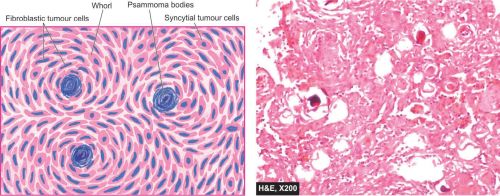
- The tumour cells have features of both syncytial and fibroblastic type forming whorls which contain central laminated areas of calcification called psammoma bodies.
Meningioma Grades
| GRADE I | GRADE II | GRADE III |
|---|---|---|
|
|
|
Immunohistochemical staining
- EMA ⊕ in ~80%