Carpal Tunnel: Difference between revisions
Jump to navigation
Jump to search
No edit summary |
|||
| Line 1: | Line 1: | ||
==Boundaries of the Carpal Tunnel== | ==Boundaries of the Carpal Tunnel== | ||
<div> | |||
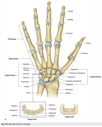
[[File:Hand and wrist joints, bones.png|thumb|252x252px]] | |||
* '''Roof''': Flexor retinaculum. | * '''Roof''': Flexor retinaculum. | ||
* '''Floor''': Carpal bones (hamate, capitate, trapezoid, trapezium, pisiform, triquetrum, lunate, and scaphoid). | * '''Floor''': Carpal bones (hamate, capitate, trapezoid, trapezium, pisiform, triquetrum, lunate, and scaphoid). | ||
* '''Medial Boundary''': Pisiform and hamate bones. | * '''Medial Boundary''': Pisiform and hamate bones. | ||
* '''Lateral Boundary''': Scaphoid and trapezium bones. | * '''Lateral Boundary''': Scaphoid and trapezium bones. | ||
</div> | |||
==Attachments of the Flexor retinaculum== | ==Attachments of the Flexor retinaculum== | ||
*'''Medially''': It attaches to the pisiform and the hook of the hamate. | *'''Medially''': It attaches to the pisiform and the hook of the hamate. | ||
*'''Laterally''': It attaches to the scaphoid tubercle and the trapezium. | *'''Laterally''': It attaches to the scaphoid tubercle and the trapezium. | ||
Revision as of 11:14, 13 September 2023
Boundaries of the Carpal Tunnel

- Roof: Flexor retinaculum.
- Floor: Carpal bones (hamate, capitate, trapezoid, trapezium, pisiform, triquetrum, lunate, and scaphoid).
- Medial Boundary: Pisiform and hamate bones.
- Lateral Boundary: Scaphoid and trapezium bones.
Attachments of the Flexor retinaculum
- Medially: It attaches to the pisiform and the hook of the hamate.
- Laterally: It attaches to the scaphoid tubercle and the trapezium.