Posterior Cerebral Artery: Difference between revisions
Jump to navigation
Jump to search
No edit summary |
|||
| Line 34: | Line 34: | ||
|} | |} | ||
= Branches of PCA = | |||
== P1 segment == | == P1 segment == | ||
<table class="wikitable" width="100%"> | <table class="wikitable" width="100%"> | ||
Revision as of 05:28, 3 August 2024
Related pages
Vascular territory

- Parietooccipital sulcus (medial) and inferior temporal sulcus (lateral)
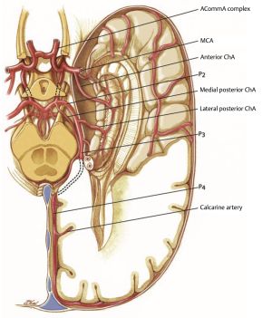
Anatomical segments
- P1: PCA from the origin to posterior communicating artery (AKA mesencephalic, precommunicating, circular, peduncular, basilar...). The long and short circumflex and thalamoperforating arteries arise from P1
- P2: PCA from origin of PComA to the origin of inferior temporal arteries (AKA ambient, post-communicating, perimesencephalic), P2 traverses the ambient cistern, hippocampal, anterior temporal, peduncular perforating, and medial posterior choroidal arteries arise from P2
- P3: PCA from the origin of the inferior temporal branches to the origin of the terminal branches (AKA quadrigeminal segment). P3 traverses the quadrigeminal cistern
- P4: segment after the origin of the parieto-occipital and calcarine arteries, includes the cortical branches of the PCA
| Segment | Description |
|---|---|
| P1 | Arises from the basilar bifurcation and extends through the interpeduncular cistern to the junction with the posterior communicating artery |
| P2A (anterior) | Runs in the crural cistern, extending from the PComA to the lateral mesencephalic sulcus where it becomes the P2P (posterior) segment |
| P2P (posterior) | Runs in the ambient cistern, lateral to the midbrain |
| P3 | Runs in the quadrigeminal cistern, separated from the P2P segment by the tectal plate |
| P4 | Begins at the calcarine sulcus |
Branches of PCA
P1 segment
| P1 Segment | Feature |
|---|---|
| Posterior thalamoperforator arteries |
|
| Medial posterior choroidal arteries |
|
| Meningeal branches | Supply: tentorium and the falx |
P2 segment
| P2 Segment | Feature |
|---|---|
| Lateral posterior choroidal artery |
|
| Thalamogeniculate arteries | Medial geniculate body, lateral geniculate body, pulvinar, superior colliculus, and crus cerebri |
| Cortical branches |
|
P3 segment
| P3 Segment | Feature |
|---|---|
| Posterior temporal artery |
|
| Internal occipital artery | |
| Parietooccipital artery |
|
| Calcarine artery |
|
| Posterior pericallosal artery |
|