Internal Carotid Artery: Difference between revisions
Jump to navigation
Jump to search
(Created page with "<h1>Bouthillier Classification of ICA Segments</h1><br><img src="https://www.fmichael1.com/moodle/images/Neurosurgery-Board-Review-QA/part1-Basic_Science/1-neuroanatomy/q17.jpg" alt="" width="565" height="575" class="img-responsive atto_image_button_text-bottom"><br><br><br> <table style="border-collapse:collapse;border-spacing:0;margin:0px auto;" class="tg"> <tbody> <tr> <td style="font-family:Arial, sans-serif;font-size:14px;padding:10px 5px;bor...") |
No edit summary |
||
| Line 1: | Line 1: | ||
<h1>Bouthillier Classification of ICA Segments</h1> | <h1>Bouthillier Classification of ICA Segments</h1> | ||
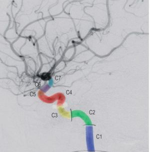
[[File:Bouthillier Classification of ICA Segments.jpg|thumb]] | |||
<table style="border-collapse:collapse;border-spacing:0;margin:0px auto;" class="tg"> | <table style="border-collapse:collapse;border-spacing:0;margin:0px auto;" class="tg"> | ||
<tbody> | <tbody> | ||
Revision as of 17:25, 17 January 2024
Bouthillier Classification of ICA Segments

| C1 (Cervical) |
|
| C2 (Petrous) |
|
| C3 (Lacerum) |
|
| C4 (Cavernous) |
|
| C5 (Clinoid) |
|
| C6 (ophthalmic/supraclinoid) |
|
| C7 (communicating) |
|