Internal Carotid Artery: Difference between revisions
Jump to navigation
Jump to search
No edit summary |
No edit summary |
||
| Line 1: | Line 1: | ||
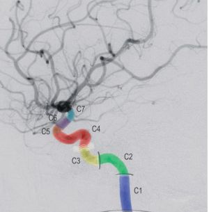
= Bouthillier Classification of ICA Segments = | |||
[[File:Bouthillier Classification of ICA Segments.jpg|thumb]] | [[File:Bouthillier Classification of ICA Segments.jpg|thumb]] | ||
== C1 (Cervical) == | |||
* Extends from origin of ICA to the [[Skull Base|skull base]] | |||
== C2 (Petrous) == | |||
* Artery location: within the carotid canal of the petrous temporal bone. | |||
* Initial movement: ascends vertically within the canal (vertical portion). | |||
* Subsequent movement: turns anteriorly, medially, and superiorly within the canal (genu). | |||
* Continued movement: continues horizontally (horizontal portion) toward the petrous apex. | |||
* Exit point: exits the temporal bone at the petrous apex. | |||
* Related arteries: Vidian artery and Caroticotympanic artery (variable). | |||
== C3 (Lacerum) == | |||
* Starting point: where the internal carotid artery exits from the carotid canal. | |||
* Extension: extends up to the level of the petroclinoid ligament. | |||
* Path: passes over (not through) the covered foramen lacerum. | |||
== C4 (Cavernous) == | |||
* Starting point: at the superior aspect of the petroclinoid ligament. | |||
* Included region: the portion of the internal carotid artery coursing through the cavernous sinus until the proximal dural ring. | |||
** Meningohypophyseal artery: arises from the posterior genu of C4 and gives rise to three major branches. | |||
** Inferior hypophyseal artery: supplies the posterior [[pituitary gland]]. | |||
** Tentorial artery (of Bernasconi and Cassinari). | |||
** Dorsal clival (meningeal) artery. | |||
* Inferolateral trunk: commonly arises from the horizontal portion of C4 and courses laterally to supply several areas. | |||
** Supplies [[cranial nerves]] III, IV, and VI. | |||
** Supplies the trigeminal ganglion. | |||
** Supplies the dura covering the cavernous sinus. | |||
== C5 (Clinoid) == | |||
* Short segment b/w the proximal & distal dural rings | |||
* Related to the anterior clinoid process | |||
== C6 (ophthalmic/supraclinoid) == | |||
* Starting point: at the distal dural ring/reflection (continuous with the falx) around the anterior clinoid process. | |||
* Considered intradural: at the point around the anterior clinoid process, it enters the subarachnoid space. | |||
* Extension: extends to the origin of the posterior communicating artery. | |||
* Ophthalmic artery: typically arises from the medial aspect of the C6 segment and courses with the optic nerve through the optic canal into the orbit. | |||
** The ophthalmic artery gives rise to multiple ocular, orbital, and extraorbital branches. | |||
** Ocular branches include the central retinal artery and ciliary arteries. | |||
* Superior hypophyseal artery: arises from the medial aspect of C6, anastomoses with its contralateral counterpart, and forms a vascular plexus about the pituitary stalk. | |||
** This plexus supplies the anterior pituitary gland, tuber cinereum, optic nerve, and optic chiasm. | |||
== C7 (communicating) == | |||
* Starting point: just proximal to the origin of the posterior communicating artery. | |||
* Termination: where the internal carotid artery bifurcates into the anterior and middle cerebral arteries. | |||
* Posterior communicating artery: courses posteriorly through the suprasellar cistern to anastomose with the PCA. | |||
** A large PCoA size suggests that it directly supplies the PCA territory as a persistent fetal PCA. | |||
** The origin of the PCoA often exhibits a focal enlargement, known as the infundibulum. | |||
** CN III courses through the suprasellar cistern close to the PCoA, making it susceptible to aneurysms of the PCoA. | |||
** The anterior thalamoperforating arteries arise from the PCoAs and supply parts of the medial hypothalamus, thalamus, and lateral aspect of the third ventricle. | |||
* Anterior choroid artery: arises from the posterior aspect of C7 just above the PCoA, and its long course is divided into three segments. | |||
** First, it courses through the suprasellar cistern just medial to the uncus of the temporal lobe (cisternal segment). | |||
** Then, it turns laterally and passes through the choroid fissure to enter the temporal horn of the lateral ventricle. | |||
** Within the ventricle, the anterior choroid artery supplies the choroid plexus and courses posterosuperiorly with the choroid plexus up to and around the pulvinar of the thalamus. | |||
** It supplies the medial temporal lobe, the optic tract and lateral geniculate body, the dorsal globus pallidus, the inferior half of the posterior limb of the internal capsule, the lateral aspect of the cerebral peduncle, the tail of the caudate nucleus, and the choroid plexus. | |||
[[Category:Neuroanatomy]] | [[Category:Neuroanatomy]] | ||
[[Category:Vascular anatomy]] | [[Category:Vascular anatomy]] | ||
Revision as of 01:09, 5 March 2024
Bouthillier Classification of ICA Segments

C1 (Cervical)
- Extends from origin of ICA to the skull base
C2 (Petrous)
- Artery location: within the carotid canal of the petrous temporal bone.
- Initial movement: ascends vertically within the canal (vertical portion).
- Subsequent movement: turns anteriorly, medially, and superiorly within the canal (genu).
- Continued movement: continues horizontally (horizontal portion) toward the petrous apex.
- Exit point: exits the temporal bone at the petrous apex.
- Related arteries: Vidian artery and Caroticotympanic artery (variable).
C3 (Lacerum)
- Starting point: where the internal carotid artery exits from the carotid canal.
- Extension: extends up to the level of the petroclinoid ligament.
- Path: passes over (not through) the covered foramen lacerum.
C4 (Cavernous)
- Starting point: at the superior aspect of the petroclinoid ligament.
- Included region: the portion of the internal carotid artery coursing through the cavernous sinus until the proximal dural ring.
- Meningohypophyseal artery: arises from the posterior genu of C4 and gives rise to three major branches.
- Inferior hypophyseal artery: supplies the posterior pituitary gland.
- Tentorial artery (of Bernasconi and Cassinari).
- Dorsal clival (meningeal) artery.
- Inferolateral trunk: commonly arises from the horizontal portion of C4 and courses laterally to supply several areas.
- Supplies cranial nerves III, IV, and VI.
- Supplies the trigeminal ganglion.
- Supplies the dura covering the cavernous sinus.
C5 (Clinoid)
- Short segment b/w the proximal & distal dural rings
- Related to the anterior clinoid process
C6 (ophthalmic/supraclinoid)
- Starting point: at the distal dural ring/reflection (continuous with the falx) around the anterior clinoid process.
- Considered intradural: at the point around the anterior clinoid process, it enters the subarachnoid space.
- Extension: extends to the origin of the posterior communicating artery.
- Ophthalmic artery: typically arises from the medial aspect of the C6 segment and courses with the optic nerve through the optic canal into the orbit.
- The ophthalmic artery gives rise to multiple ocular, orbital, and extraorbital branches.
- Ocular branches include the central retinal artery and ciliary arteries.
- Superior hypophyseal artery: arises from the medial aspect of C6, anastomoses with its contralateral counterpart, and forms a vascular plexus about the pituitary stalk.
- This plexus supplies the anterior pituitary gland, tuber cinereum, optic nerve, and optic chiasm.
C7 (communicating)
- Starting point: just proximal to the origin of the posterior communicating artery.
- Termination: where the internal carotid artery bifurcates into the anterior and middle cerebral arteries.
- Posterior communicating artery: courses posteriorly through the suprasellar cistern to anastomose with the PCA.
- A large PCoA size suggests that it directly supplies the PCA territory as a persistent fetal PCA.
- The origin of the PCoA often exhibits a focal enlargement, known as the infundibulum.
- CN III courses through the suprasellar cistern close to the PCoA, making it susceptible to aneurysms of the PCoA.
- The anterior thalamoperforating arteries arise from the PCoAs and supply parts of the medial hypothalamus, thalamus, and lateral aspect of the third ventricle.
- Anterior choroid artery: arises from the posterior aspect of C7 just above the PCoA, and its long course is divided into three segments.
- First, it courses through the suprasellar cistern just medial to the uncus of the temporal lobe (cisternal segment).
- Then, it turns laterally and passes through the choroid fissure to enter the temporal horn of the lateral ventricle.
- Within the ventricle, the anterior choroid artery supplies the choroid plexus and courses posterosuperiorly with the choroid plexus up to and around the pulvinar of the thalamus.
- It supplies the medial temporal lobe, the optic tract and lateral geniculate body, the dorsal globus pallidus, the inferior half of the posterior limb of the internal capsule, the lateral aspect of the cerebral peduncle, the tail of the caudate nucleus, and the choroid plexus.