Spine Disorders in Children: Difference between revisions
Jump to navigation
Jump to search
(Created page with "thumb|419x419px {| class="wikitable" !Malformation Type !<u>Type I (diastematomyelia)</u> !'''Type II (diplomyelia)''' |- |Anatomic Features |Two hemicords in two dural sleeves separated by a midline bony spur. |Two hemicords in a single dural sleeve. Hemicords are separated and tethered by a fibrous band attached to the dura. |- |Radiographic Features |Bony spur seen on CT. CT features of intersegmental fus...") |
No edit summary |
||
| Line 50: | Line 50: | ||
* Chiari malformation (6–7%) | * Chiari malformation (6–7%) | ||
|} | |} | ||
[[Category:Pediatric Neurosurgery]] | |||
Revision as of 19:38, 2 March 2024

| Malformation Type | Type I (diastematomyelia) | Type II (diplomyelia) |
|---|---|---|
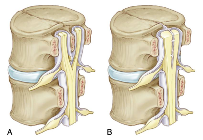
| Anatomic Features | Two hemicords in two dural sleeves separated by a midline bony spur. | Two hemicords in a single dural sleeve. Hemicords are separated and tethered by a fibrous band attached to the dura. |
| Radiographic Features | Bony spur seen on CT. CT features of intersegmental fusion and adjacent spina bifida. MRI shows two hemicords in separate subarachnoid spaces. | Two hemicords seen within a single subarachnoid space on T2WI. ± fibrous septum attached to the dura. |
| Location | Typically lumbar | May occur anywhere along the spinal axis |
| Symptoms | Cutaneous Markers | Associated Findings on Px | Associated Anomalie |
|---|---|---|---|
|
|
|
|