Anterior Inferior Cerebellar Artery: Difference between revisions
Jump to navigation
Jump to search
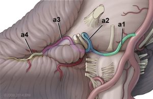
(Created page with "The Anterior Inferior Cerebellar Arteries (AICAs) typically arise from the lower half of the basilar artery (BA), most commonly from a single trunk. The AICA is divided into four segments: the anterior pontine (a1), the lateral pontine (a2), the flocculopeduncular (a3), and the cortical (a4) segments. * The A1 segment extends from its origin to the midpoint of the inferior olive. * The A2 segment extends from the inferior olive to the flocculus and includes several branc...") |
No edit summary |
||
| Line 1: | Line 1: | ||
[[File:AICA segments.jpg|thumb]] | |||
The Anterior Inferior Cerebellar Arteries (AICAs) typically arise from the lower half of the basilar artery (BA), most commonly from a single trunk. | The Anterior Inferior Cerebellar Arteries (AICAs) typically arise from the lower half of the basilar artery (BA), most commonly from a single trunk. | ||
The AICA is divided into four segments: the anterior pontine (a1), the lateral pontine (a2), the flocculopeduncular (a3), and the cortical (a4) segments. | The AICA is divided into four segments: the anterior pontine (a1), the lateral pontine (a2), the flocculopeduncular (a3), and the cortical (a4) segments. | ||
| Line 5: | Line 6: | ||
* The A3 segment extends from the flocculus to the cerebellopontine fissure. | * The A3 segment extends from the flocculus to the cerebellopontine fissure. | ||
* The A4 segment is the portion of the vessel distal to the cerebellopontine fissure. | * The A4 segment is the portion of the vessel distal to the cerebellopontine fissure. | ||
[[Category:Neuroanatomy]] | [[Category:Neuroanatomy]] | ||
[[Category:Vascular anatomy]] | [[Category:Vascular anatomy]] | ||
Latest revision as of 23:15, 4 March 2024

The Anterior Inferior Cerebellar Arteries (AICAs) typically arise from the lower half of the basilar artery (BA), most commonly from a single trunk. The AICA is divided into four segments: the anterior pontine (a1), the lateral pontine (a2), the flocculopeduncular (a3), and the cortical (a4) segments.
- The A1 segment extends from its origin to the midpoint of the inferior olive.
- The A2 segment extends from the inferior olive to the flocculus and includes several branches: the labyrinthine, the subarcuate, the cerebellosubarcuate, and recurrent perforating arteries.
- The A3 segment extends from the flocculus to the cerebellopontine fissure.
- The A4 segment is the portion of the vessel distal to the cerebellopontine fissure.