Arachnoid cysts
Introduction
AKA leptomeningeal cysts, distinct from posttraumatic leptomeningeal cysts (AKA growing skull fractures), and unrelated to infection.
Arachnoid cysts (AC) are congenital lesions that arise during development from splitting of arachnoid membrane (thus they are technically intra-arachnoid cysts) and contain fluid that is usually identical to CSF. They do not communicate with the ventricles or subarachnoid space.
May be unloculated or may have septations.
Typically lined with meningothelial cells positive for epithelial membrane antigen (EMA) and negative for carcinoembryonic antigen (CEA). AC may also occur in the spinal canal.
“Temporal lobe agenesis syndrome” is a label that had been used to describe the findings with middle cranial fossa ACs.
This term is now obsolete since brain volumes on each side are actually the same, bone expansion and shift of brain matter account for the parenchyma that appears to be replaced by the AC.
Two types of histological findings:
- “simple arachnoid cysts”: arachnoid lining with cells that appear to be capable of active CSF secretion. Middle fossa cysts seem to be exclusively of this type
- cysts with more complex lining which may also contain neuroglia, ependyma, and other tissue types
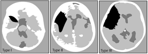
Galassi classification

- Type I: small, biconvex, located in anterior temporal tip. No mass effect. Communicates with subarachnoid space on water-soluble contrast CT cisternogram (WS-CTC).
- Type II: involves proximal and intermediate segments of Sylvian fissure. Completely open insula gives rectangular shape. Partial communication on WS-CTC.
- Type III: involves entire Sylvian fissure. Marked midline shift. Bony expansion of middle fossa (elevation of lesser wing of sphenoid, outward expansion of squamous temporal bone). Minimal communication on WS-CTC. Surgical treatment usually does not result in total reexpansion of brain (may approach type II lesion).