Cranial Dysraphisms
Key concepts
- Cranial dysraphisms range in severity from minimally symptomatic dermal sinus tracts to large encephaloceles.
- The overall incidence of encephaloceles is declining, possibly due to dietary folate supplementation.
- Encephaloceles can occur in various sites and exhibit variation in size, shape, and contents.
- Prenatal diagnosis of encephaloceles is common, typically confirmed by elevated α-fetoprotein levels and ultrasound.
- Surgical repair of extreme herniation may not provide significant benefits and requires counseling and consultation.
- Surgical treatment aims to reduce herniation, preserve viable brain, and reconstruct craniofacial defects.
- Prognosis varies based on factors specific to the location and anatomy of the encephalocele.
- Cranial meningoceles can occur in diverse locations, and complete excision with primary dural closure leads to a good prognosis.
- Dermal sinus tracts present as cutaneous dimples and are often associated with a cyst. Total en bloc resection is the management goal.
- Complete resections of dermal sinus tracts have a favorable prognosis and low recurrence rates.
NOSOLOGY (Classification and Terminology)
- Cephalocele: Refers to the herniation of any intracranial contents through a skull defect.
- Encephalocele: Contains normal brain tissue.
- Gliocele: Contains gliotic tissue.
- Cranial meningocele: Contains only cerebrospinal fluid (CSF) surrounded by arachnoid tissue.
- Encephalocystocele: Refers to a herniated sac that contains ventricular tissue.
- Atretic cephalocele: Considered a forme fruste encephalocele characterized by an involuted cephalocele with a small, noncystic, flat lesion located in the midline of the scalp.
- Cranium bifidum: Represents a rare nonunion of cranial sutures, usually with the brain covered by dura and pericranium, and intact overlying skin. Small foramina may be a normal variant and can be familial, persisting in up to 60% of the population.
- Exencephaly: Refers to the development of a substantial portion of the brain in a patient with acrania, representing the most extreme end of cranial dysraphism.
Diagnosis
Maternal Serum α-Fetoprotein (MSAFP)
- MSAFP ↑ in early 2nd trimester (optimal: 16-18 weeks, extended: 14-21 weeks) often used for initial NTD screen in routine prenatal care.
- MSAFP level 2.5x median at 16-18 weeks → 98% chance of open NTD; found in 79% of pregnancies w/ open NTDs & 3% of nml singleton pregnancies.
- Single MSAFP sampling gives Dx accuracy of 60-70%; if predicted risk > 1 in 500, repeat MSAFP ✓ recommended.
- Constant or ↓ MSAFP on repeat testing → NTD likely not present; 40% of initially ↑ MSAFP return to normal levels on repeat testing.
- Persistent ↑ MSAFP or initial ↑ MSAFP → high-resolution fetal ultrasonography recommended.
- False-positive MSAFP ↑ can be due to incorrect gestational age, multiple pregnancy, fetal demise, or other fetal abnormalities (e.g., omphalocele, cloacal exstrophy, esophageal atresia, annular pancreas, duodenal atresia, gastroschisis, congenital nephrosis, polycystic kidneys, urinary tract obstruction, renal agenesis).
- If MSAFP > 3x mean on initial & repeat testing, but fetal ultrasound is normal → repeat ultrasonography recommended.
High-Resolution Fetal Ultrasonography (HRFU)

- Screens for NTDs ~100% accuracy; predicts anatomic level of spinal cord defect 64% of the time & within one level 79% of the time
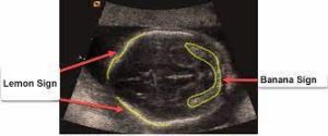
- Often visualizes two cranial abnormalities assoc. w/ hydrocephalus & Chiari II malformations:
- Lemon sign: scalloping of frontal bones on biparietal view, present in 80% of fetuses w/ myelomeningocele
- Banana sign: abnormal midbrain shape, elongated cerebellum, & obliteration of the cisterna magna, present in 93% of myelomeningocele fetuses; false-positive rate only 0.88%
- Banana Sign נצפה בדרך כלל לפני שבוע 26 להריון ונמצא ביותר מ-90% מהמקרים של ההפרעה הזו
- Both signs highly diagnostic of myelomeningocele, even when spinal abnormality not directly visualized
- If ultrasonography non-diagnostic, MRI or amniocentesis recommended
Youmans Video
Video 213 1 Cranial Dysraphisms
                   <source src="https://www.fmichael1.com/moodle/local/VOD_gal/media/youmans8e/VOLUME_2/SECTION_8_Pediatrics/PART_2_Cranial_Development_Abnormalities/Video_213_1_Cranial_Dysraphisms.mp4" type="video/mp4">
                   <track default="" kind="captions" label="English" src="https://www.fmichael1.com/moodle/local/VOD_gal/media/youmans8e/VOLUME_2/SECTION_8_Pediatrics/PART_2_Cranial_Development_Abnormalities/Video_213_1_Cranial_Dysraphisms.vtt" srclang="en">
</video>