Spine Disorders in Children

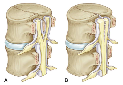
| Malformation Type | Type I (diastematomyelia) | Type II (diplomyelia) |
|---|---|---|
| Anatomic Features | Two hemicords in two dural sleeves separated by a midline bony spur. | Two hemicords in a single dural sleeve. Hemicords are separated and tethered by a fibrous band attached to the dura. |
| Radiographic Features | Bony spur seen on CT. CT features of intersegmental fusion and adjacent spina bifida. MRI shows two hemicords in separate subarachnoid spaces. | Two hemicords seen within a single subarachnoid space on T2WI. ± fibrous septum attached to the dura. |
| Location | Typically lumbar | May occur anywhere along the spinal axis |
| Symptoms | Cutaneous Markers | Associated Findings on Px | Associated Anomalie |
|---|---|---|---|
|
|
|
|