Synaptic Transmission
Overview
- The human brain consists of 86 billion neurons.
- Communication among these neurons is facilitated by synapses.
Types of Synapses
Two main types: Electrical and Chemical Synapses.
- Electrical synapses use connexons for direct current flow.
- Direct, passive flow of electrical current.
- Chemical synapses use neurotransmitters for cell-to-cell communication.
- Chemical: Communication via neurotransmitter release.
| Feature | Electrical Synapses | Chemical Synapses |
|---|---|---|
| Structure |
|
|
| Function |
|
|
| Key Characteristics |
|
|
- Electrical and chemical synapses differ fundamentally in their transmission mechanisms
-
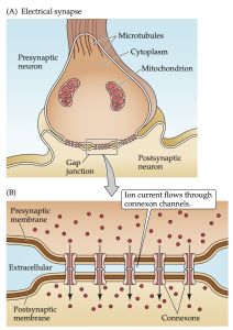
(A) At electrical synapses, gap junctions occur between pre- and postsynaptic membranes. (B) Gap junctions contain connexon channels that permit current to flow passively from the presynaptic cell to the postsynaptic cell.
-
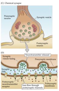
(C) At chemical synapses, there is no intercellular continuity, and thus no direct flow of current from pre- to postsynaptic cell. (D) Synaptic current flows across the postsynaptic membrane only in response to the secretion of neurotransmitters, which open or close postsynaptic ion channels after binding to receptor molecules on the postsynaptic membrane.


Overview of Chemical Synapses
- Presynaptic terminal with synaptic vesicles.
- Postsynaptic cell, separated by synaptic cleft.
- Filamentous elements in pre- and postsynaptic processes.
- Structures in synaptic cleft, including active zone and postsynaptic density.
[[
|thumb|Overview of postsynaptic signaling ]]
Chemical Synapses and Neurotransmitters
- Over 100 different neurotransmitters identified.
- Communication process: Synthesis, packaging, release, and binding of neurotransmitters, followed by rapid removal or degradation.
Signaling Transmission Sequence at Chemical Synapse

- Synaptic Vesicle Preparation → ormation and filling of synaptic vesicles with neurotransmitter.
- Action Potential Arrival - Invades presynaptic terminal → Opens voltage-gated Ca2+ channels.
- Ca2+ Influx:
- Rapid influx due to concentration gradient (10–3 M external vs. 10–7 M internal).
- Elevates Ca2+ in presynaptic terminal.
Exocytosis and Postsynaptic Response
- Exocytosis
- Ca2+ elevation → Synaptic vesicles fuse with presynaptic membrane.
- Neurotransmitters released into synaptic cleft.
- Neurotransmitter Binding
- Diffuses across cleft → Binds to postsynaptic receptors.
- Opens/closes channels in postsynaptic membrane, altering ion flow.
- Postsynaptic Neuron Response
- Alters conductance and membrane potential.
- Increases/decreases probability of firing an action potential.
Termination of Signal
- Neurotransmitter Removal
- Uptake into glial cells or enzymatic degradation.
- Effect
- Terminates neurotransmitter action.
- Ensures transient information transmission from one neuron to another.
Synaptic Transmission at the Neuromuscular Junction (NMJ)

- NMJ: Connection between alpha-motor neuron axons and skeletal muscle fibers.
- Mechanism: Cholinergic transmission using acetylcholine (Ach).
- Relevance: Basics of neurotransmitter release applicable across all synapses.
Sequence of Events in Synaptic Transmission
- Action Potential Arrival: Depolarizes presynaptic membrane → opens voltage-gated Ca2+ channels.
- Ca2+ Influx: Triggers release of Ach.
- Ach Release and Binding: Binds to nicotinic receptor on muscle membrane → depolarization (EPP).
- Muscle Action Potential: Triggered by EPP → muscle contraction.
- Ach Breakdown: Acetylcholinesterase (AchE) terminates Ach action → reuptake of choline.
Other Cholinergic Synapses
- Neuronal Nicotinic (NN) Receptors: In autonomic ganglia, activated by presynaptic cholinergic neurons.
- Muscarinic Receptors: In parasympathetic target tissues, G-protein coupled receptors.
Synapses Between Neurons
- Location: On the cell body and dendrites.
- Influence: Closer synapses to the axon hillock have greater influence on action potential initiation.
- EPSP vs. IPSP: EPSP increases excitability (more likely to fire), while IPSP decreases excitability (less likely to fire).
- Key Receptors: EPSP (Nicotinic, Non-NMDA, NMDA), IPSP (GABAA&C, Glycine).
EPSP and IPSP Explained
- EPSP: Increased Na+ conductance, similar to EPP at NMJ.
- IPSP: Increased Cl- conductance, reducing neuron's excitability.
- Important Receptors:
- EPSP: Nicotinic, Non-NMDA, NMDA (ligands: Ach, glutamate, aspartate).
- IPSP: GABAA&C, Glycine (ligands: GABA, glycine).