Malformations of Cortical Development

From NeuroWiki
Jump to navigation Jump to search

Categories

  • group I: abnormal cell proliferation or apoptosis
  • group II: abnormal neuronal migration
  • group III: abnormal cortical organization
Group Characteristics Conditions
Group I abnormal proliferation of neuronal and glial cells
  • Microcephaly
  • macrocephaly
  • FCDs without and with balloon cells (Taylor dysplasia types IIa and IIb),
  • hemimegalencephaly,
  • tuberous sclerosis,
  • dysembryoplastic neuroepithelial tumors (DNETs), gangliogliomas, gangliocytomas
Group II abnormal neuronal migration
  • Lissencephaly
  • Band Heterotopia
Group III abnormal cortical organization
  • Polymicrogyria,
  • schizencephaly,
  • type I FCDs,
  • mild MCDs


Group Affected step of development MCDs resulting from the disturbance Short definition of the MCD
Group I Progenitor cell proliferation and apoptosis Microcephaly Abnormally small head and brain
Macrocephaly Abnormally big head and brain
Hemimegalencephaly Overgrowth of (part of) a cerebral hemisphere
Focal cortical dysplasia Disturbed lamination and dysmorphic neurons
Group II Neuronal migration Lissencephaly type I Absence of normal convolutions/folds
Periventricular heterotopia (PH) Neurons accumulating at the ventricles underneath a normal cortex
Subcortical band heterotopia/double cortex Band of grey matter located between the lateral ventricular wall and the cortex
Group III Neuronal organisation Cobblestone lissencephaly/lissencephaly type II Overmigration of neurons to localize on the surface of a brain with reduced gyri
Polymicrogyria Too many (usually small) folds/convolutions
Schizencephaly Fluid-filled cleft from ventricle(s) to pia lined by heterotopic grey matter

Group II - Neuronal migration disorders
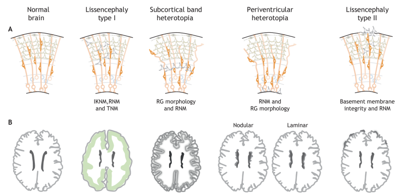
(A) Schematics highlighting the cellular basis of NMDs in the adult human cortex, showing the different cortical layers and neuronal migrations. Single ectopic neurons are shown in grey and the affected structures or processes are indicated. (B) Schematic showing the MRI-detectable morphological defects in the adult human brain that are caused by the cellular defects of each NMD. Ectopically located clusters of affected neurons are shown as greyshading. Lissencephaly type I is characterized bya smooth brain surface and a simplified fourlayered cortex (indicated by green shading). In subcortical band heterotopia, the cortex contains an additional band of grey matter underneath the white matter. Periventricular heterotopia is characterized by clusters (nodular) or sheets (laminar) of neurons accumulating at the ventricles underneath a normal cortex. In lissencephaly type II, neuronsovermigrate ontothecorticalsurface. Schematicsareadapted fromMRIimages,seee.g.BizzottoandFrancis,2015;Francisetal., 2006; Guerrini and Parrini, 2010. IKNM, interkinetic nuclear migration; RG, radial glia; RNM, radial neuronal migration; TNM, tangential neuronal migration.