Vidian nerve
Jump to navigation
Jump to search


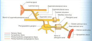
- Greater & deep petrosal nerves → vidian nerve; traverses vidian canal, unites w/ maxillary nerve & pterygopalatine ganglion in pterygopalatine fossa.
- Vidian nerve sends branches to zygomatic nerve → anastomosis w/ lacrimal nerve to gland.
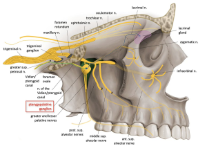
- Pterygopalatine fossa: contains maxillary nerve branches, vidian nerve junction w/ pterygopalatine ganglion, & terminal branches of maxillary artery.
- Vidian nerve enters posterior margin of sphenopalatine ganglion in pterygopalatine fossa via vidian canal.
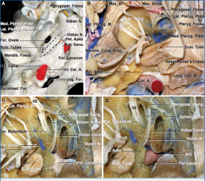
- Foramen rotundum (for maxillary nerve) & pterygoid canal (for vidian nerve) open through posterior wall of fossa formed by sphenoid pterygoid process.
- Vidian nerve penetrates upper part of pterygoid process & area below foramen rotundum to enter pterygopalatine fossa.
- Vidian nerve: union of greater petrosal nerve (parasympathetic fibers from facial nerve @ geniculate ganglion level) & deep petrosal nerve (sympathetic fibers from carotid plexus) → lacrimal gland & nasal mucosa.
- Pterygoid canal (inferomedial to foramen rotundum) carries vidian nerve (autonomic fibers) to pterygopalatine ganglion.
- Pterygoid plates contain pterygoid canal (for vidian nerve), form lateral wall of choana medially, pterygopalatine fossa at center, & pterygomaxillary fissure laterally.
- Vidian nerve key for ICA location in transclival drilling.
- Vidian nerve indicates ICA as it turns up @ foramen lacerum (lacerum seg).
- Transpyterygoid approach: uncinate process removal, anterior & posterior ethmoid cells opening, wide sphenoidotomy; vidian nerve & sphenopalatine ganglion carefully displaced laterally before anterior pterygoid process removal.
- Pterygopalatine fossa contents: vidian nerve & artery, pterygopalatine ganglion & branches, & maxillary nerve & artery and branches.
- Preserve ophthalmic branch (V1) of trigeminal nerve in dissection, vidian nerve likely damaged in transpterygopalatine fossa approach, especially if vidian canal guides to petrous ICA.